Suisse

INDIVIDUAL HIP®

Individualized hip stem

LEARN MOREeast
keyboard_arrow_down

The reference in individualized products

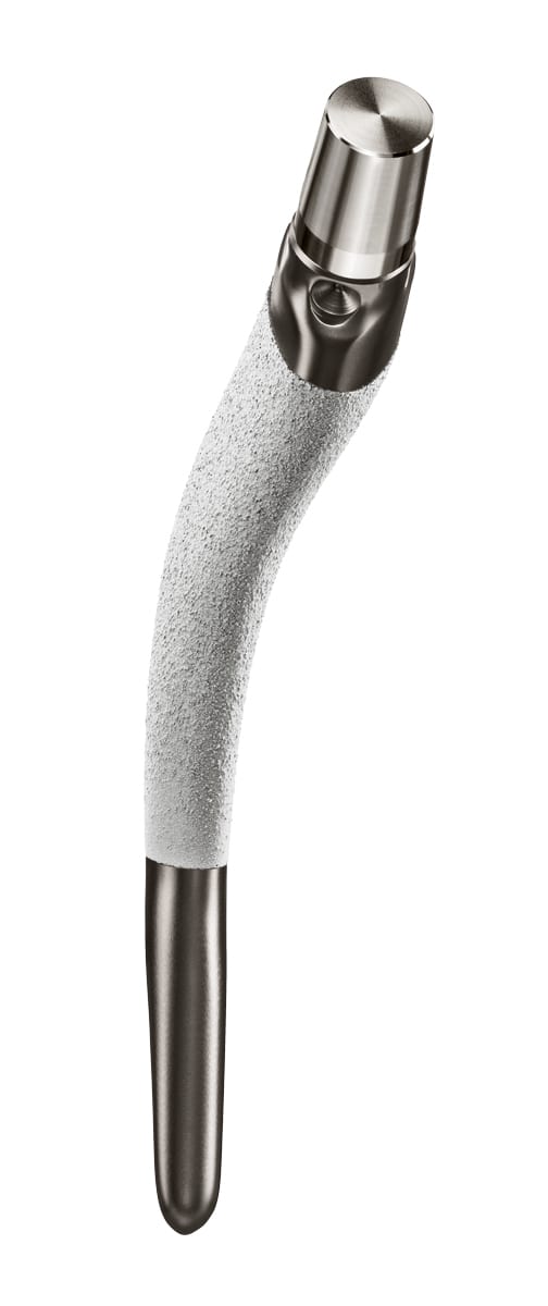
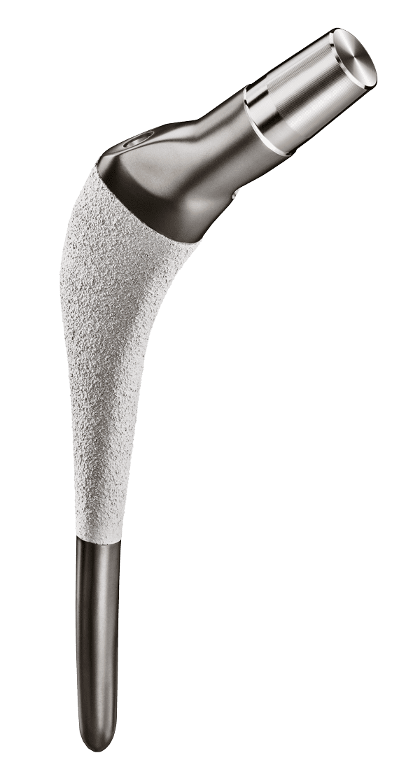
With more than 25,000 individualized-designed stems, and over 30 years’ clinical experience, the INDIVIDUAL HIP® stem is the benchmark in individualized hip replacement.

20 years

Clinical experience of the concept, and 25 years of published knowledge (1)

96.8%

20-year survival for patients under 50 years of age (1)(2)

Individualized

to replicate the proximal femur

Every patient is unique

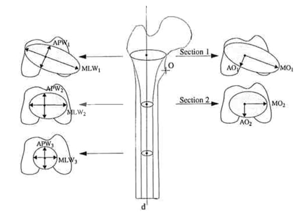
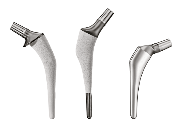
Femoral morphology varies greatly from one person to another in the three dimensions of space. Research conducted by Husmann (3) and Sariali (4) has demonstrated these variations in the proximal femur and hip in 3D, even in cases often considered “normal”. In some rarer cases, the hip anatomy can be dysplastic and deviate greatly from the anatomical averages for which standard hip prostheses were designed.

No-compromise femur reproduction

Each individual is different, so the INDIVIDUAL HIP® stem is designed to adapt perfectly to the shape of the medullary canal in all three dimensions, so no need for compromise:


  • Supports designed to adapt most effectively to the mediolateral flare of the intramedullary canal;
  • Anatomic sagittal curvature reproducing the anterior curvature of the patient’s femur;
  • Restoration of the natural torsion of the femur on the axial plane.

Metaphyseal fixation

Symbios’ engineers identify the preferred support zones between the bone and the prosthesis within the metaphyseal zone to:


  • maximise primary mechanical stability regardless of the size and shape of the canal;
  • ensure proximal load transmission to promote bone remodelling and osseointegration.

Compaction of cancellous bone

A individualized compactor identical to the implant is delivered for each patient, to prepare the medullary canal before implanting the final stem.


  • Compaction of cancellous bone, to preserve good intramedullary vascularisation for better postoperative osteogenesis;
  • Individualized smooth compactor adapting to the shape of the intramedullary canal;
  • Optional pricked rasp to make it easier to work with denser bones.

Individualized

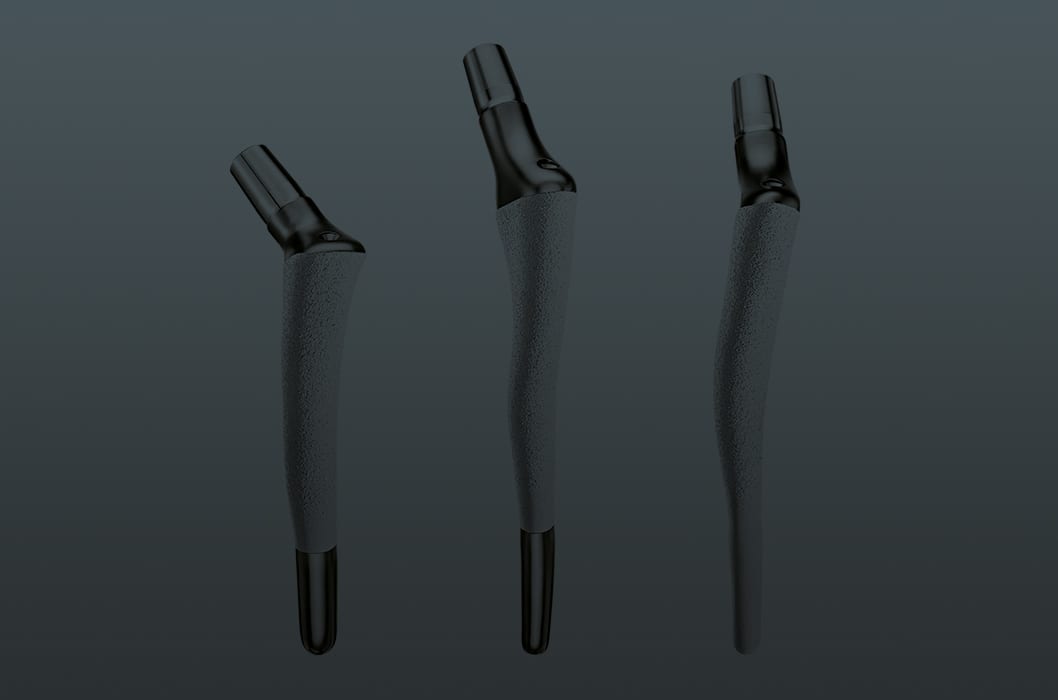
for no-compromise hip reconstruction

Complexity of the femoral anatomy

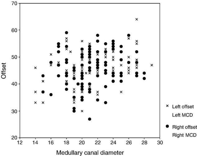
Work undertaken by Krishnan (5) has shown that there is no correlation between medullary canal size and femoral offset. Standard implants often become limited because of their homothetic design when the femoral offset to be restored is not proportional to the size of the canal.

No-compromise reconstruction

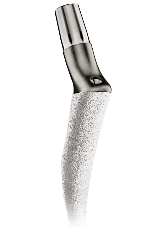
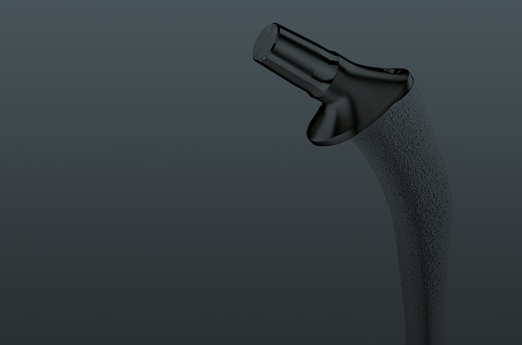
The individualized stem has a neck designed to reconstruct the joint by restoring the centre of rotation with no compromise based on the planned stem position.


  • Adjustment of CCD angle and neck length;
  • Restoration of leg length and femoral offset, regardless of the size and shape of the stem;
  • Adjustment of prosthetic anteversion.

Restored femoral anteversion

The stem is designed to restore femoral anteversion in a personalised manner and to maximise anterior-posterior stability and joint function.


  • Restoration of native anteversion or correction of anteversion according to the planning agreed between the surgeon and the Symbios engineer;
  • Consideration of the patient’s step angle, allowing the lower limb to function efficiently.

Individualized

to accommodate surgical preferences

A multitude of options

In addition to adapting to the patient, the individualized stem adapts to the standard practices of each surgeon, with a multitude of options and variants:


  • Cementless individualized stem
    • Optional full or partial stem coating;
    • Optional collar.
  • Individualized cemented stem
    • Recommended when the primary stability criteria for a cementless option are not met;
    • Optional collar.
  • Individualized smooth compactor
    • For optimal compaction of the spongious bone.
  • Individualized pricked rasp
    • To make working with very dense bone easier.

Preoperative planning

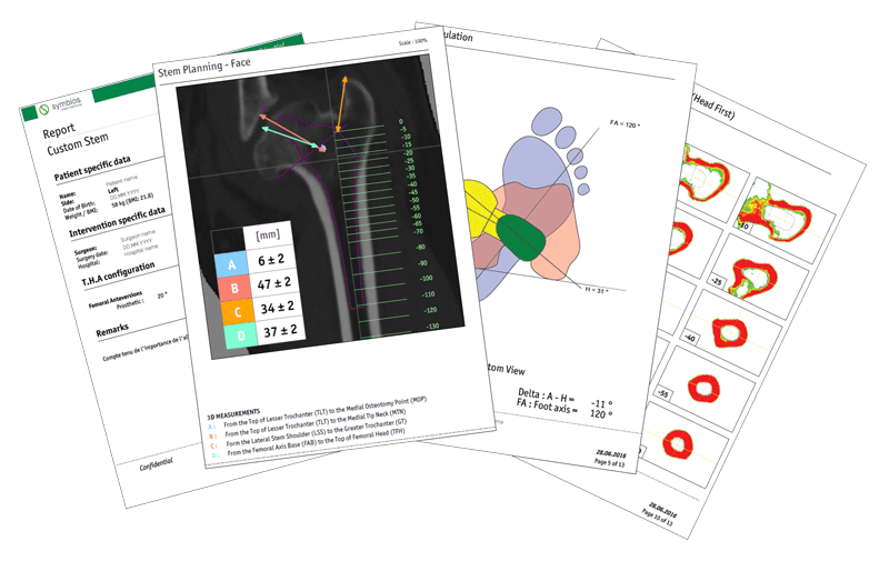
The INDIVIDUAL HIP® stem comes with the planning report, to guide the surgeon throughout the procedure.


  • Reminder of the preoperative analysis (leg lengths, acetabular and femoral geometries, densities, etc.);
  • Guide for neck resection, cup positioning and preparing the femoral canal;
  • Numerous checks available to control the position of implants during surgery.

Resources to download

(1) Dessyn E, Flecher X, Parratte S, Ollivier M, Argenson JN. A 20-year follow-up evaluation of total hip arthroplasty in patients younger than 50 using a custom cementless stem. Hip Int. 2019 Sep ;29(5) :481-488.

(2) Flecher X, Pearce O, Parratte S, Aubaniac JM, Argenson JN. Custom cementless stem improves hip function in young patients at 15-year followup. Clin Orthop Relat Res. 2010 Mar;468(3):747-55.

(3) Husmann O, Rubin P J, Leyvraz PF, De Roguin B, Argenson JN. Three-dimensional morphology of the proximal femur. J Arthroplasty 1997 Jun ;12(4):444-50.

(4) Sariali E, Mouttet A, Pasquier G, Durante E. Three-dimensional hip anatomy in osteoarthritis. Analysis of the femoral offset. J. Arthoplasty 2009 Sep ;24(6):990-7.

(5) Krishnan SP, Carrington RW, Mohiyaddin S, Garlick N. Common misconceptions of normal hip joint relations on pelvic radiographs. J Arthroplasty. 2006 Apr;21(3):409-12.

(6) Argenson JN, Flecher X, Parratte S, Aubaniac JM. Anatomy of the dysplastic hip and consequences for total hip arthroplasty. Clin Orthop Relat Res. 2007 Dec;465:40-5.

(7) Flecher X, Parratte S, Aubaniac JM, Argenson JN. Three-dimensional custom-designed cementless femoral stem for osteoarthritis secondary to congenital dislocation of the hip. J Bone Joint Surg Br. 2007 Dec;89(12):1586-91.

(8) Argenson JN, Ryembault E, Flecher X, Brassart N, Parratte S, Aubaniac JM. Three-dimensional anatomy of the hip in osteoarthritis after developmental dysplasia. J Bone Joint Surg Br. 2005 Sep;87(9):1192-6.

(9) Flecher X, Parratte S, Aubaniac JM, Argenson JN. Cementless total hip arthroplasty using custom stem and reinforcement ring in hip osteoarthritis following developmental dysplasia. Hip Int. 2007;17 Suppl 5:S120-7.

(10) Wettstein M, Mouhsine E, Argenson JN, Rubin PJ, Aubaniac JM, Leyvraz PF. Three-dimensional computed cementless custom femoral stems in young patients: midterm followup. Clin Orthop Relat Res. 2005 Aug;(437):169-75.

(11) Koulouvaris P, Stafylas K, Xenakis T. Cementless modular centroid reconstruction cup in young adults with congenital dysplasia of the hip. J Arthroplasty. 2008 Jan;23(1):79-85.

(12) Koulouvaris P, Stafylas K, Sculco T, Xenakis T. Custom-design implants for severe distorted proximal anatomy of the femur in young adults followed for 4-8 years. Acta Orthop. 2008 Apr;79(2):203-10.

(13) Flecher X, Parratte S, Brassart N, Aubaniac JM, Argenson JN. Evaluation of the hip center in total hip arthroplasty for old developmental dysplasia. J Arthroplasty. 2008 Dec;23(8):1189-96.

(14) Flecher X, Ollivier M, Maman P, Pesenti S, Parratte S, Argenson JN. Long-term results of custom cementless stem total hip arthroplasty performed in hip fusion. Int Orthop. 2018 Jun;42(6):1259-1264.

(15) Flecher X, Blanc G, Sainsous B, Parratte S, Argenson JN. A customised collared polished stem may reduce the complication rate of impaction grafting in revision hip surgery: a 12-year follow-up study. J Bone Joint Surg Br. 2012 May;94(5):609-14.

(16) Rubin PJ, Leyvraz PF, Aubaniac JM, Argenson JN, Estève P, De Roguin B. The morphology of the proximal femur. A three-dimensional radiographic analysis. J Bone Joint Surg Br. 1992 Jan ;74(1):28-32.

(17) Wettstein M, Mouhsine E, Argenson JN, Rubin PJ, Aubaniac JM, Leyvraz PF. Three-dimensional computed cementless custom femoral stems in young patients: midterm followup. Clin Orthop Relat Res. 2005 Aug;(437):169-75. doi: 10.1097/01.blo.0000163001.14420.3a. PMID: 16056046.